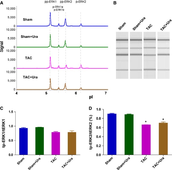
Fig. 3.

Urantide doesn*t affect the phosphorylation of extracellular signal-regulated kinas (ERK) in cardiac side population cells (CSPs) in pressure overload mice. (A) Multiple isoforms of Phospho-ERK was detected in CSPs by nanofluidic proteomic immunoassay (NIA). Peaks on the traces that represent phosphorylated isoforms are indicated. (B) NIA pseudoblot representation of ERK. (C) Bar graph of tp-ERK1/tERK1 by NIA quantification. tp-ERK1: total phosphorylation of ERK1. tERK1: total ERK1. (D) Bar graph of tp-ERK2/ERK2 by NIA quantification. tp-ERK2: total phosphorylation of ERK2. tERK2: total ERK2. Urantide (30 μg/kg/day) or vehicle were respectively continuously administered by Alzet osmotic minipumps to mice from 2 to 4 weeks after transverse aorta constriction (TAC) or sham operation, then CSPs were isolated from heart by fluorescence-activated cell sorting for NIA with antibody to phosphos-ERK. Values are expressed as mean ± SEM. Sham:n = 9. Sham+Ura:n = 6. TAC:n = 6. TAC+Ura:n = 7. Ura:urantide. *P < 0.05 versus sham mice.
