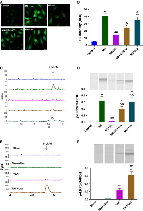
Fig. 5.

UII inhibits the phosphorylation of LRP6 in cardiac side population cells (CSPs) by UT/JNK signaling during mechanical stretch. (A) Immunofluorescent images revealed that positive signal (green) could be observed in cultured CSPs with antibody to p-LRP6 (1:200); bar: 20 μm. (B) Quantitative evaluation of p-LRP6 by analysis of fluorescence intensity. (C) P-LRP6 was detected in cultured CSPs by nanofluidic proteomic immunoassay (NIA). Peaks on the traces that represent phosphorylated isoforms of LRP6 are indicated. (D) NIA pseudoblot representation of p-LRP6 and bar graph of p-LRP6/GAPDH in cultured CSPs by NIA quantification. Cultured CSPs were pre-treated with PBS, urantide (Ura, 1 μM) or SP600125 (SP6, 5 μM), respectively, for 30 min, then subjected to mechanical stretch (MS) and incubated with PBS or UII (0.1 μM) for 3 hrs. Values are expressed as mean ± SEM. **P < 0.01 versus control; ##P < 0.01 versus control; #P < 0.05 versus MS; &P < 0.05 versus MS plus UII; &&P < 0.01 versus MS plus UII. The experiment was repeated for at least three times. (E) P-LRP6 was detected in CSPs isolated from transverse aorta constriction (TAC) or Sham mice by NIA. (F) NIA pseudoblot representation of p-LRP6 and bar graph of p-LRP6/GAPDH in isolated CSPs by NIA quantification. Values are expressed as mean ± SEM. Sham:n = 9; Sham+Ura:n = 6; TAC:n = 6; TAC+Ura:n = 7. Ura: urantide. **P < 0.01 versus Sham mice; ##P < 0.01 versus TAC mice.
