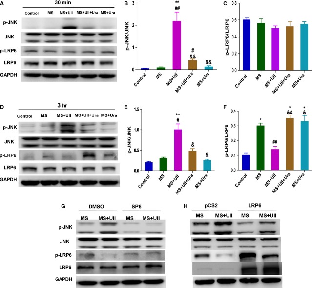
Fig. 6.
UII inhibits the phosphorylation of LRP6 in UT cell line by activating JNK during mechanical stretch. Western blot analysis for p-LRP6 and p-JNK levels. UT cells were pre-treated with Urantide (Ura, 1 μM) for 30 min., then subjected to MS and treated with UII for 30 min. and 3 hrs. (A) Representative picture of p-LRP6 and p-JNK level at 30 min. after UII treatment during MS. Quantitative evaluation of p-JNK (B) and p-LRP6 (C) by analysis of Western blot. (D) Representative picture of p-LRP6 and p-JNK level at 3 hrs after UII treatment during MS. Quantitative evaluation of p-JNK (E) and p-LRP6 (F) by analysis of Western blot. P-JNK and p-LRP6 were analysed in UT cells pre-treated with SP600125 (SP6, 5 μM; G) or overexpressed LRP6 (H) with or without UII (0.1 μM) during MS. UT cells were pre-treated with SP600125 or DMSO for 30 min., then subjected to MS and treated with UII for 3 hrs. UT cells were overexpressed with LRP6-pCS2 or pCS2 for 48 hrs, then subjected to MS and treated with UII for 3 hrs. Values are expressed as mean ± SEM. *P < 0.05, **P < 0.01 versus control. #P < 0.05, ##P < 0.01 versus MS. &P < 0.05, &&P < 0.01 versus MS plus UII. The experiment was repeated for at least three times.

