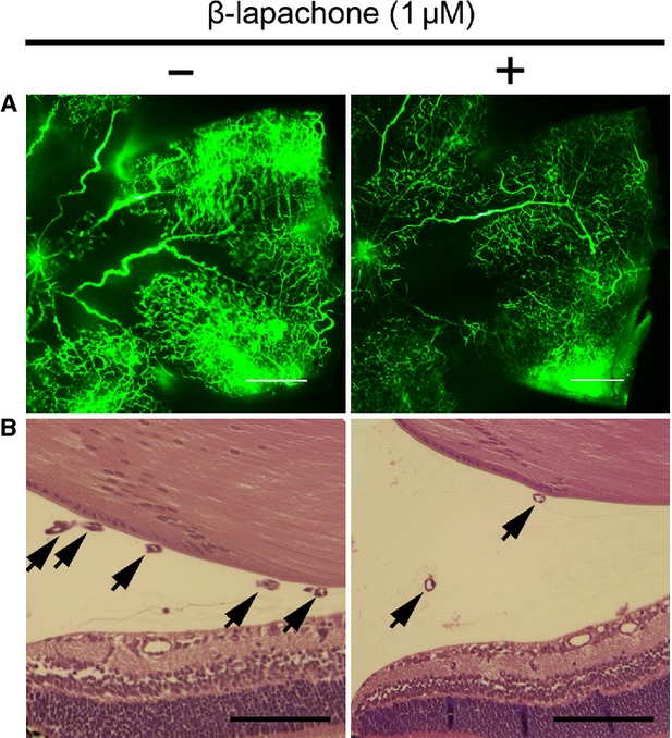
Fig. 1.

Intraocular injection of β-lapachone inhibits retinal neovascularization in oxygen-induced retinopathy (OIR). (A) Retinal vasculatures in control and β-lapachone–treated mice with OIR were evaluated with fluorescein angiography. Whole mount retinal preparation from P17 control and 1 μM β-lapachone intravitreously injected mice of OIR were performed after 1-hr perfusion of fluorescein-conjugated dextran (MW = 500,000). Neovascular tufts of intravitreous neovascularization were observed at the border of vascular and avascular retina; scale bar: 500 μm. (B) Vascular lumen was counted for quantitative analysis of retinal neovascularization in OIR. Haematoxylin and eosin–stained cross-sections were prepared from P17 control and 1 μM β-lapachone–treated mice of OIR. Arrows indicate the vascular lumens of new vessels growing into the vitreous. Number of vascular lumens was counted from randomly selected ×40 magnification view; scale bar: 100 μm.
