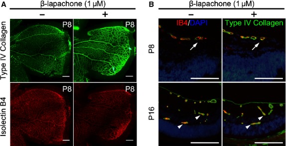
Fig. 4.

β-lapachone never mitigates physiological retinal angiogenesis in the retinal development. After intraocular injection of 1 μM β-lapachone or 0.1% dimethyl sulfoxide in 1 μl PBS on P4, the effect of β-lapachone on physiological retinal angiogenesis in the developmental retina was evaluated at P8 and P16. (A) For primary vascular plexus, whole mount retinal preparation were stained with type IV collagen (green) and isolectin B4 (red); scale bars: 200 μm. (B) Retinal cross-sections at P8 and P16 were also evaluated. Retinal vessels were immunostained with type IV collagen (green) and isolectin B4 (red), and nucleus were counterstained with DAPI (blue). Physiological superficial vascular plexus was not regressed by β-lapachone in the peripheral retina at P8. Physiological retinal angiogenesis to intermediate and deep vascular plexus was not affected by β-lapachone at P16. Arrows indicate superficial vessels reaching to the peripheral retina. Arrowheads indicate deep and intermediated plexus; scale bars: 200 μm.
