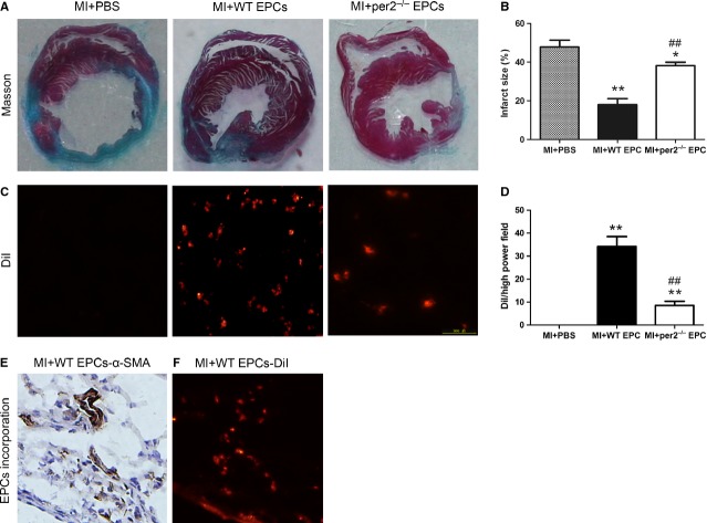
Fig. 5.
Effect of endothelial progenitor cells (EPCs) with or without per2 on mouse infarct area 4 weeks after myocardial infarction (MI). (A) Representative infarct area in the three groups and (B) quantification. (C) Representative expression of DiI-labelled EPCs in the myocardium and (D) quantification (*P < 0.05, **P < 0.01 versus MI+PBS, ##P < 0.01 versus MI+wild-type EPCs; n = 6). (E) α-Smooth muscle actin (α-SMA) expression in mouse cardiac tissues. (F) DiI-labelled EPC expression in similar locations.

