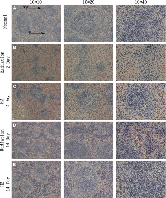
Fig. 2.

Morphology of spleen after in vivo γ-radiation. (A) Spleens of mice with control morphology are comprised of both red and white pulps. (B) Spleens of mice 2 days after in vivo γ-radiation. (C) Spleens in mice pre-treated with H2 at 2 days after irradiation. (D) Spleens of mice 14 days after in vivo γ-radiation. (E) Spleens in mice pre-treated with H2 at 14 days after irradiation.
