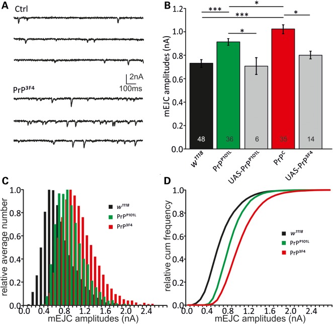
Figure 2.
Prion causes enlarged miniature EJCs. TEVC recordings were performed at the NMJ to investigate the effects of neuronal PrP3F4 or PrPP101L expression. (A) Sample traces showing mEJCs in control (Ctrl [w1118]) and PrP3F4 expressing larvae. (B) Mean mEJC amplitudes are increased following PrP3F4 and PrPP101L expression with PrPP101L showing a reduction relative to PrP3F4 (n—number of NMJs is indicated in bars. ***P < 0.001, *P < 0.05, n = 6–48 NMJs, [n = 19 animals for w1118, n = 15 animals for PrPP101L, n = 16 animals for PrP PrP3F4, n = 4–8 animals for UAS controls]). Data denote mean ± SEM, ANOVA with Tukey–Kramer post hoc test. (C) Normalized amplitude histograms for mEJCs show a right-shift of amplitudes following PrP3F4 expression. PrPP101L expression left-shifted amplitudes relative to PrPC but still caused an increase versus control (w1118). There is no evidence for multi-quantal release as distributions do not exhibit multiple peaks. (D) Relative cumulative amplitude histograms illustrate the right-shift in amplitudes for PrP3F4 expressing larvae. PrPP101L expression induced a left-shift relative to PrP3F4 (K–S test, PrP3F4 versus w1118: D = 0.22, P = 0.024, PrPP101L versus w1118: D = 0.30, P = 0.00023, PrP3F4 versus PrPP101L: D = 0.42, P < 0.00001). Distributions for UAS controls are not shown for simplicity.

