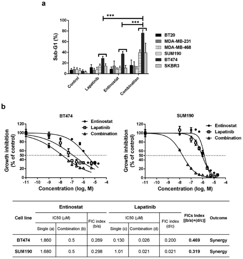
Fig. 1.

The combination of entinostat and lapatinib inhibited proliferation of HER2+ breast cancer cells. a Cells (2×10⁵) were placed on a 6-well plate overnight, treated with or without the drugs (entinostat, 5.0 μM for all cell lines; lapatinib, 1.0 μM for HER2+ cell lines and 5.0 μM for HER2- cell lines) for 72 hours, and then stained with propidium iodide (PI) for cell cycle analysis using flow cytometry. Each bar represents the mean of 3 independent experiments; error bars, SD. *** p <0.001 combination compared with either entinostat or lapatinib. b Cells were treated with entinostat or lapatinib or both in combination for 72 hours. For combination, entinostat was fixed with 0.5 μM and mixed with various range of lapatinib (0.01 – 20 μM). And then a WST-1 proliferation assay was performed. For analysis, the non-linear fit curve method was used via GraphPad Prism software. The table represents the synergistic inhibitory effect of entinostat and lapatinib. The fractional inhibitory concentration (FIC) for the combination is the sum of the FICs of the 2 drugs and was interpreted as follows: < 0.5, synergy; 0.5 – 2.0, additive; > 2.0, antagonistic.
