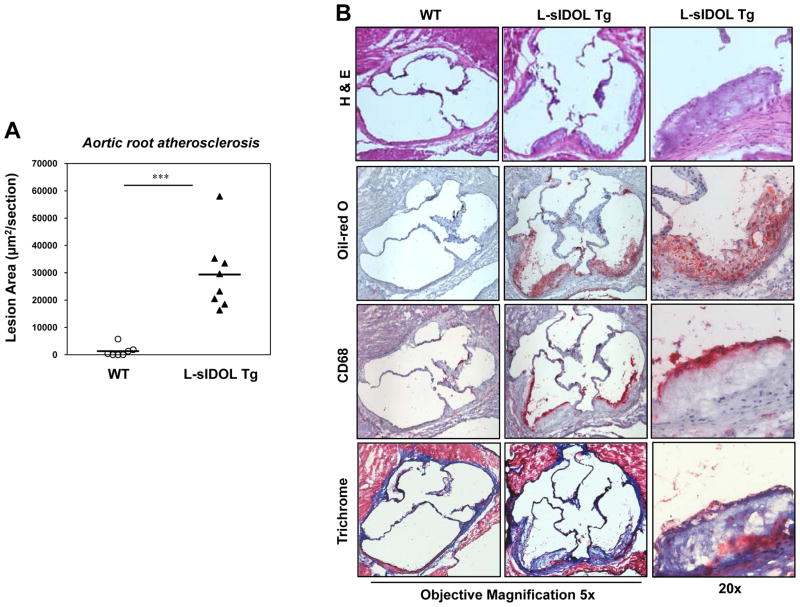
Figure 5. L-sIDOL mice develop atherosclerosis in the aortic root.
(A) Quantitation of aortic root lesions in male mice following 30 weeks of western diet; (B) Representative pictures of aortic root lesions in WT and L-sIDOL mice stained with haemotoxylin and eosin (H&E; top), Oil red O (top middle), CD68 (bottom middle) and trichrome (bottom) at magnifications as indicated.

