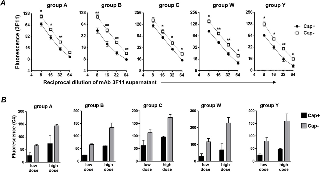
Fig. 6.
Capsule limits binding of and C4b deposition by anti-LOS IgM mAb 3F11. A. Binding of mAb 3F11 to Cap+ and Cap− isogenic meningococcal mutants. Bacteria were incubated with varying dilutions (X-axis) of tissue culture supernatant containing mAb 3F11 and 3F11 binding was measured by flow cytometry (shown as median fluorescence on the log2 Y-axis; mean of 3 experiments). Binding to Cap+ is shown by the solid line and binding to Cap− by the broken line. B. All 5 capsular groups limit anti-LOS IgM-mediated C4 deposition. Cap+ (black bars) and Cap− (grey bars) isogenic mutants were incubated with low (1/32 and 1/64 for Cap+ and Cap− mutants, respectively) or high (1/8 and 1/16 for Cap+ and Cap− mutants, respectively) concentrations of 3F11 supernatants. Two-fold higher concentrations of 3F11 were used on Cap+ mutants as a conservative estimate to normalize for differences in 3F11 binding as shown in panel A. C4 deposition was measured by FACS following the addition of purified C1 complex and C4. Median fluorescence of C4b binding was recorded and each bar shows the mean (range) of two independent experiments. Supplemental Figure S3 shows representative histograms of C4b deposition.

