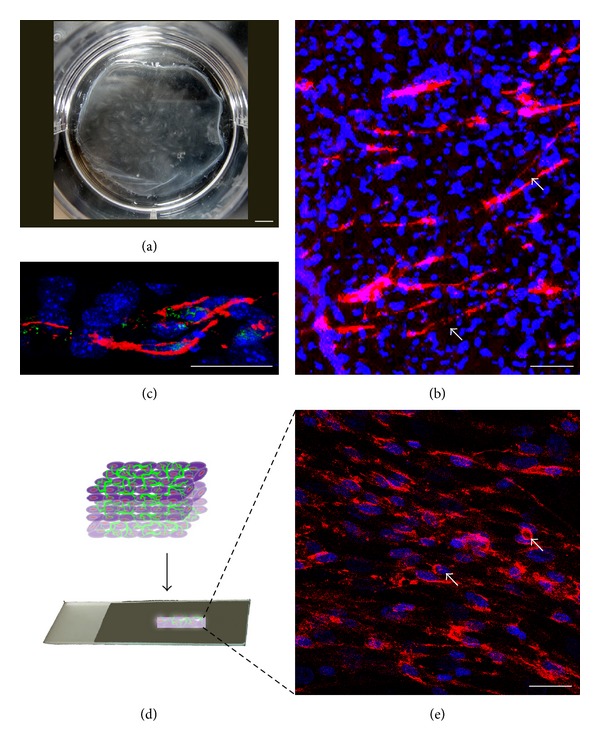
Figure 3.

This picture shows the overall view of a cell sheet (a) (Scale bar = 3.4 mm). Confocal microscopy images show the lumens formation of HUVECs on hMSCs sheet after 7 days of culture. Immunofluorescent staining images of CD31 show lumens formed on the hMSCs sheet (b) (Scale bar = 100 μm) and migrated into the sheet (c) (cross section view, Scale bar = 50 μm). The three-dimensional prevascularized construct made by folding a single cell sheet was cryosectioned (d) and CD31 staining shows that networks and lumens formed in the construct (e) (white arrows, Scale bar = 100 μm).
