Figure 4.

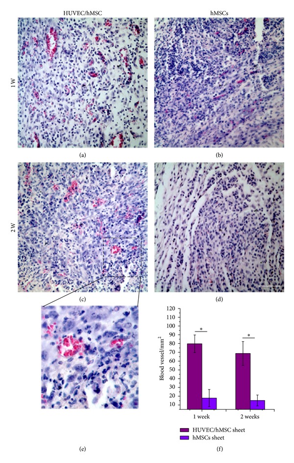
H&E staining of implants in vivo. H&E staining reveals that a large number of blood vessels grew into the HUVEC/hMSC constructs at 1 week (a) and 2 weeks (c), but few blood vessels were observed in hMSCs constructs ((b), (d)). A magnified image from (c) shows murine blood cells in a human derived blood vessel lumen (e). Blood vessel densities of the two groups at 1 and 2 weeks (f) (*P < 0.05, n = 6) (Scale bar = 100 μm).
