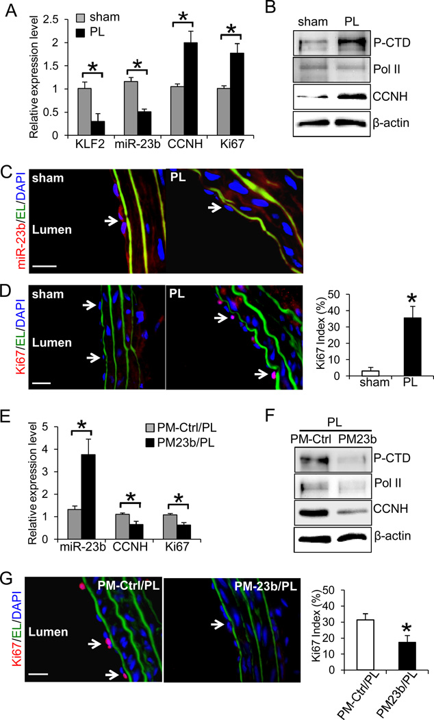
Figure 6. Flow disturbance reduces miR-23b expression and promotes EC proliferation in vivo.
A. The expression levels of miR-23b, KLF2, CCNH, and Ki67 in rat left carotid arteries (LCAs) with partial ligation (PL) or sham surgery (sham) were determined with qRT-PCR. Endothelial RNA samples were extracted from LCA by perfusing TRIzol through the carotid intima (n=4 each, *P<0.05: PL vs. sham). B. Total intima proteins were extracted from LCAs of PL and sham, and the levels of CTD phosphorylation, Pol II and CCNH were determined by Western blot. C. The decrease in miR-23b in PL was confirmed by FISH staining (red) in frozen sections. D. The increase in Ki67 in PL was demonstrated by immunofluorescence staining of Ki67 (red) in frozen sections. The Ki67 index: the number of Ki67-positive ECs / the number of ECs (n=4 each, 10 sections per sample, *P<0.05 vs. sham). Effects of PM23b treatment on E. the expression levels of miR-23b, CCNH, and Ki67 in intimal RNA extracted from PL LCAs with local delivery of PM-Ctrl (PM-Ctrl/PL) or PM23b (PM23b/PL) (n=4 each, *P<0.05: PM-Ctrl/PL vs. PM23b/PL; and F. the levels of CTD phosphorylation, Pol II, and CCNH in PM-Ctrl/PL and PM23b/PL were determined by Western blot. G. PM23b treatment reduced Ki67 staining in PL. EL: elastic lamina (green), DAPI: nuclear staining (blue). Ki67 index (n=4 each, 10 sections per sample, *P<0.05 vs. PM-Ctrl/PL). White arrows indicate the positive staining of miR-23b or Ki67 in endothelium. Results in B–D and F–G are representative images from four animals with similar results. Scale bar, 20 um.

