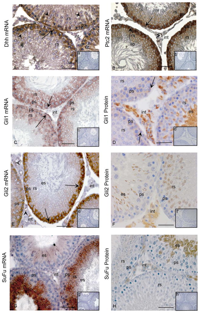
Figure 1. Cellular localization of Hedgehog signalling components in adult rat testes.
In situ hybridization with anti-sense cRNAs (A–C, E, G) and immunohistochemistry (D, F, H) were used to detect mRNAs and proteins within cells of the adult testis. Insets in each panel indicate sense controls (a′-c′,e′,g′) and controls lacking primary antibody (D, F, H). (A) Dhh mRNA in Sertoli cells (arrow heads), spermatogonia (arrow) and spermatocytes (ps) (A, anti-sense; a′ [inset], sense control). (B) Ptc2 mRNA in spermatogonia (arrows), pachytene spermatocytes (ps) and round spermatids (rs) (B, anti-sense; b′, sense). (C) Gli1 mRNA in spermatogonia (arrows), pachytene spermatocytes (ps) and round spermatids (rs) (C, anti-sense; c′ [inset], sense control). (D) Gli1 protein in spermatogonia (arrows), spermatocytes (ps), elongating spermatids (es) and some interstitial cells (int) (D, positive staining; d′ [inset], negative control). (E) Gli2 mRNA in spermatogonia (arrows) and pachytene spermatocytes (ps), endothelial cells (arrow heads) (E, anti-sense; e′ [inset], sense control). (F), Gli2 protein in round spermatids (rs), elongating spermatids (es) and interstitial cells (int) (F, positive staining; f′ [inset], negative control) (G) SuFu mRNA in round spermatids (rs) and elongating spermatids (es) (G, anti-sense; g′ [inset], sense control). (H) SuFu protein in round spermatids (rs), elongating spermatids (es) and interstitial cells (int) (H, positive staining; h′ [inset], negative control). Bars indicate 50 μm. All analyses were conducted on at least three independent specimens and yielded consistent results, with representative images shown. Positive signals are brown; all sections have blue counterstain from Hemotoxylin.

