
Figure 1. Low dose rapamycin induces functional CD8 T cell cytokine defects during LCMV infection.
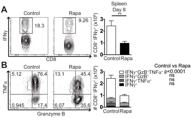
Mice were infected with 104 pfu LCMV i.p. On day 8 post-infection splenocytes were stimulated ex vivo with gp33 peptide. (A) CD8 T cells were first gated through IFNγ+ as shown in representative flow plots and quantified. (B) IFNγ+ cells were then further divided by TNFα and Granzyme B production as depicted and quantified. Representative of 2 independent repeats, each with n=8 mice/group. Unpaired two-tailed student’s t-test (A), or 2-way ANOVA (B) were used to determine statistical significance. In (A) and (C) control group is represented by open bars; rapa group is represented by filled bars. In (B) white part of the bar graph denotes the number of IFNγ+TNFα+Granzyme B+ CD8 T cells in the spleen; light grey is the number of IFNγ+Granzyme B+; dark grey is IFNγ+TNFα+; and black shows cells that are only IFNγ+.