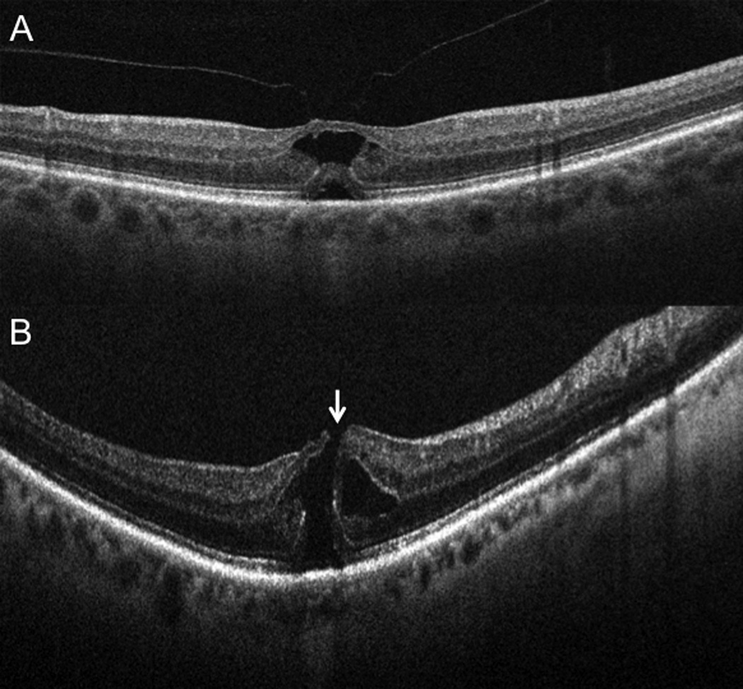
Figure 3.

Subclinical interval development of a full-thickness macular hole. (A) Preoperative 6-mm B-scan (Cirrus) at time of surgical scheduling in clinic revealing vitreomacular traction without full-thickness defect (B) Intraoperative OCT 10-mm B-scan (Bioptigen) showing interval development of subclinical full-thickness macular hole immediately prior to vitrectomy (arrow). This altered surgical approach with gas tamponade and internal limiting membrane peeling.
