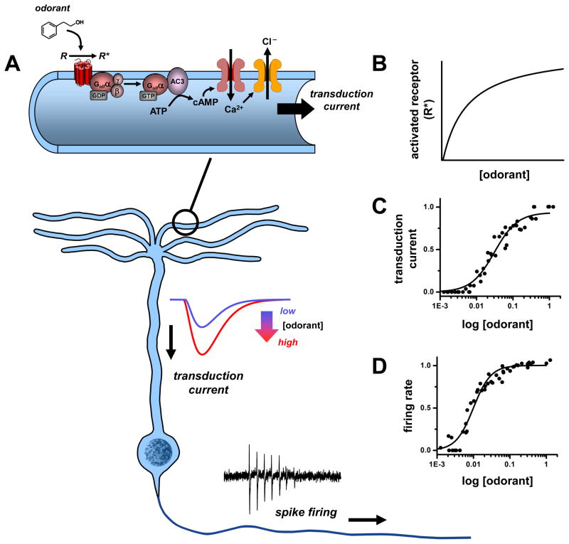
Figure 1. Odorant concentration coding in olfactory sensory neurons (OSNs).
During sensory transduction (A), odorant molecules bind and stabilize the active states of olfactory receptors (R) in ciliary membranes of OSNs. The activated receptors (R*) couple to G-proteins (Golf) and increase synthesis of cyclic AMP (cAMP) by type III adenylyl cyclase (AC3). The cAMP opens cyclic nucleotide-gated channels that conduct calcium ions into the cilia and in turn open a channel (ANO2) mediating a depolarizing efflux of chloride ions. The resulting transduction current is passed to the OSN cell body where it drives a train of action potentials (spikes). The concentration of detected odorant is encoded non-linearly at each step of transduction: by a hyperbolic dependence on the number of activated receptors (R*) in the cilia (B), a strongly cooperative variation in amplitude of the transduction current (C), and similar sigmoidal variation of spike firing rate relayed by OSN axons (D). Data source: C, D: [115], normalized currents and firing rates of frog OSN response to cineole; mammalian OSNs exhibit similar dose-response profiles.

