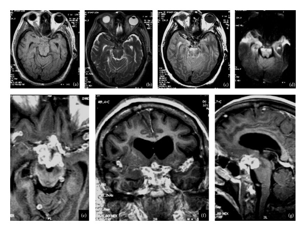
Figure 4.

(a) Axial T1-weighted images, (b) axial T2-weighted images, and (c) axial T2- and T2-FLAIR-weighted images after contrast media administration. (e) Axial plane, (f) coronal, (g) sagittal MRI images of 57-year-old woman, six years before she had tuberculous meningitis, obstructive hydrocephalus secondary to multiple coalescent nodular well-defined images; the biggest was localized in the sella, the lesion in contact with the chiasm, and extends to cavernous sinuses. Multiple lesions were localized in basal cisterns and both lateral fissures and the retrosellar extension of the lesions cause brain stem compression.
