Fig. 2.
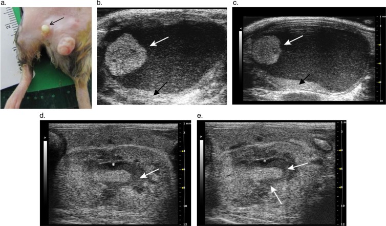
Hydronephrosis in a mouse with a preputial gland abscess and pyelonephritis. (a) Purulent discharge from a preputial gland abscess (arrow) (b) and (c). 2-D transverse images through the urinary bladder showing a round hyperechoic mass attached to the lateral bladder wall (white arrows). Echogenic debris floats in the bladder lumen and accumulates above the dorsal wall of the bladder (black arrows). The bladder wall is also thickened. In (c), the prostate gland is prominent underneath the bladder and a dilated accessory sex gland is visible dorsolateral to the bladder. (d) and (e). Longitudinal 2-D images of the left kidney with pyelonephritis, pyonephrosis, and hydronephrosis. The urine expanding the renal pelvis appears anechoic on the ultrasound image (asterisk); however, the pelvis also contains echogenic debris (arrows) due to the development of pyonephrosis in the infected kidney. The corticomedullary junction is not visible in the kidney, and there is overall patchy and increased echogenicity within both the cortex and medulla. The renal border adjacent to the dilated pelvis is irregular, due to the erosion of the renal parenchyma.

