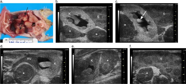
Fig. 4.
Images from a C3H/HeJ mouse with histiocytic sarcoma. (a) Gross pathologic lesions in the C3H/HeJ mouse with histiocytic sarcoma showing widespread lymphadenopathy. Tan nodules (lymph nodes) are seen effacing the kidneys (arrows) and the multiple enlarged tan lymph nodes are prominent throughout the caudal abdomen. (b) 2-D ultrasound image through the left kidney and spleen showing neoplastic infiltration of the spleen (S), kidney (K) and lymph nodes (asterisk) by histiocytic sarcoma. The renal pelvis is dilated, and the renal parenchyma has homogenous and diffusely increased echogenicity. The lymph nodes (asterisk) and spleen (S) are enlarged with prominent multifocal dark nodules scattered throughout the interior of the spleen. (c) Another view of the left kidney flanked between the spleen (S) and a large lymph node (asterisk). A section of papilla is visible showing intensely hyperechoic foci consistent with the histological finding of osseous metaplasia (arrow). (d) 2-D transverse image of the left kidney shows renal pelvic dilation and tumor thrombosis of the renal vein. The lumen of the renal vein (RV) is solid, producing gray echoes of similar echogenicity to the enlarged lymph node (asterisk) due to suspected infiltration of the vessel with neoplastic cells. The infiltrated renal vein is traceable along its length from the hilum to the inferior vena cava (IVC) and is displaced ventrally by the enlarged lymph node (asterisk). (e) The right kidney (K), and a severely enlarged perirenal lymph node (asterisk) that is distorting the cranial pole of the kidney. The right kidney also exhibits mild renal pelvic dilation. (f) Splenomegaly, with multifocal hypoechoic nodules.

