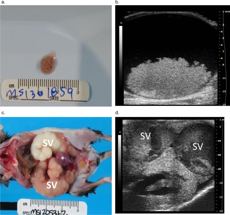
Fig. 5.
Images from male C57BL/6J mice with mouse urologic syndrome. (a) A coagulum of blood and proteinaceous material in the bladder of a mouse with bilaterally enlarged hemorrhagic seminal vesicles. The material originated from the seminal vesicles and caused an acute, severe bladder outlet obstruction. (b) The transverse 2-D image of the bladder shown in (a) shows the obstructive seminal vesicle coagulum in the bladder lumen. The large (6 mm×12 mm) hyperechoic mass is clearly visible on the floor of the bladder. (c) Urinary tract and reproductive gland lesions in a second male mouse, including enlarged tan and white seminal vesicles (SV) filled with seminal gland secretions and a hemorrhagic urinary bladder (asterisk). (d) A corresponding transverse 2-D ultrasound image for (c) shows the severely distended, sacculated seminal vesicle (SV) containing echogenic material overlying the left hydronephrotic kidney.

