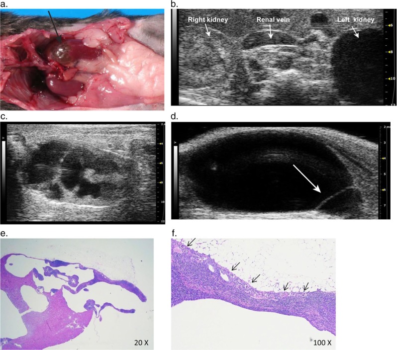
Fig. 7.
Severe unilateral hydronephrosis in the left kidney. (a) The cranial pole of the left kidney (arrow) is thin walled and appears translucent due to severe urine distension and attenuation of renal parenchyma from hydronephrosis. (b) Transverse 2-D ultrasound image showing the unaffected right kidney compared to the thin-shelled fluid filled cranial pole of the left kidney that is completely void of detectable cortical tissue. (c) 2-D longitudinal image of the left kidney with severe renal pelvic dilation, and renal parenchymal atrophy with distortion and loss of the normal renal architecture. (d) Longitudinal image of the urinary bladder showing a thin walled cystic structure (arrow) protruding into the base of the bladder, consistent with an ureterocele. (e) Histological image showing the hydronephrotic portion of the cranial pole of the kidney. (f) Histological image showing multiple small glomeruli (arrows) along the periphery of the affected cortex with an associated moderate lymphocytic interstitial infiltrate.

