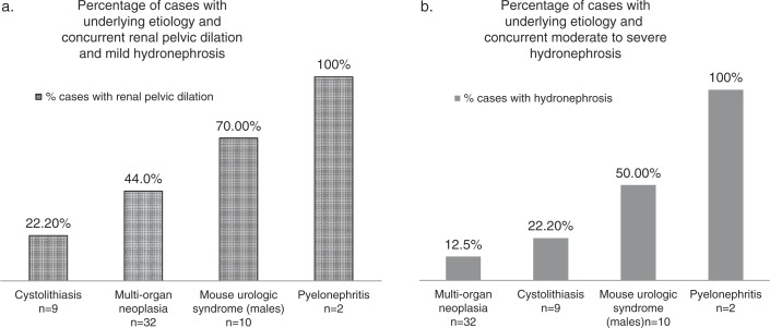
Fig. 9.
Results from the retrospective review of imaging exams and pathology reports from 156 mice aged 10–36 months. (a) Percentages of cases for designated etiologies with concurrent renal pelvic dilation and mild hydronephrosis observed on ultrasound (expanded renal pelvic space measured>14% of total diameter of kidney from transverse image near renal hilum). (b) Percentages of cases for designated etiologies with concurrent unilateral or bilateral moderate to severe hydronephrosis diagnosed at imaging (renal pelvis space expanded>30% of total width of kidney in transverse image and with significant renal parenchymal loss).

