Figure 1.
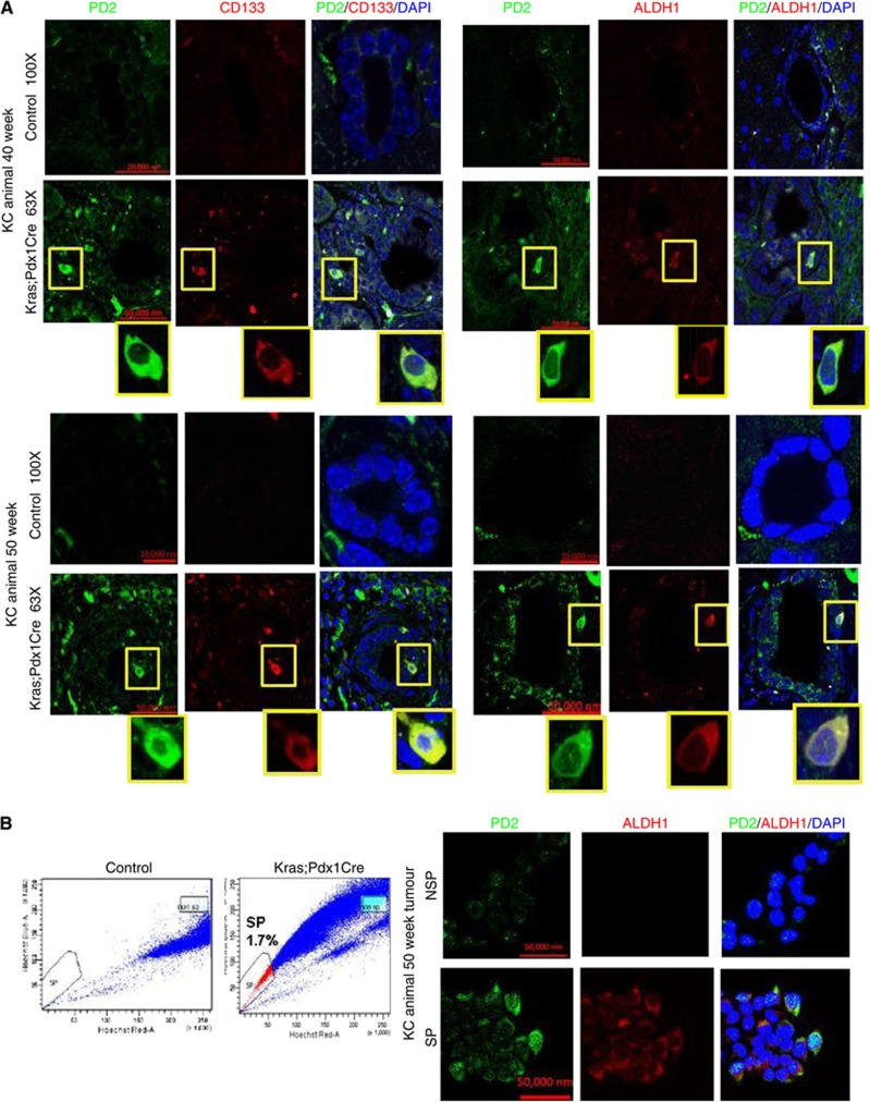
Expression of PD2 in double-transgenic mouse pancreatic cancer samples. (A) Overexpression of PD2 along with CD133 and ALDH1 in the 40th and 50th weeks of Kras-driven mouse pancreatic tumours. The control pancreas showed no expression in ductal cells. The highlighted box shows the zoomed image of single-cell staining. (B) FACS analysis was performed using Hoechst 33342 staining in Kras-driven mouse PC-driven cells. Kras;Pdx1Cre animal cells showed 1.7% of SP cells compared with control animals. Confocal images showed the overexpression of PD2 along with ALDH1 in isolated SP cells compared with NSP cells. DAPI was used as a nuclear counter staining.

