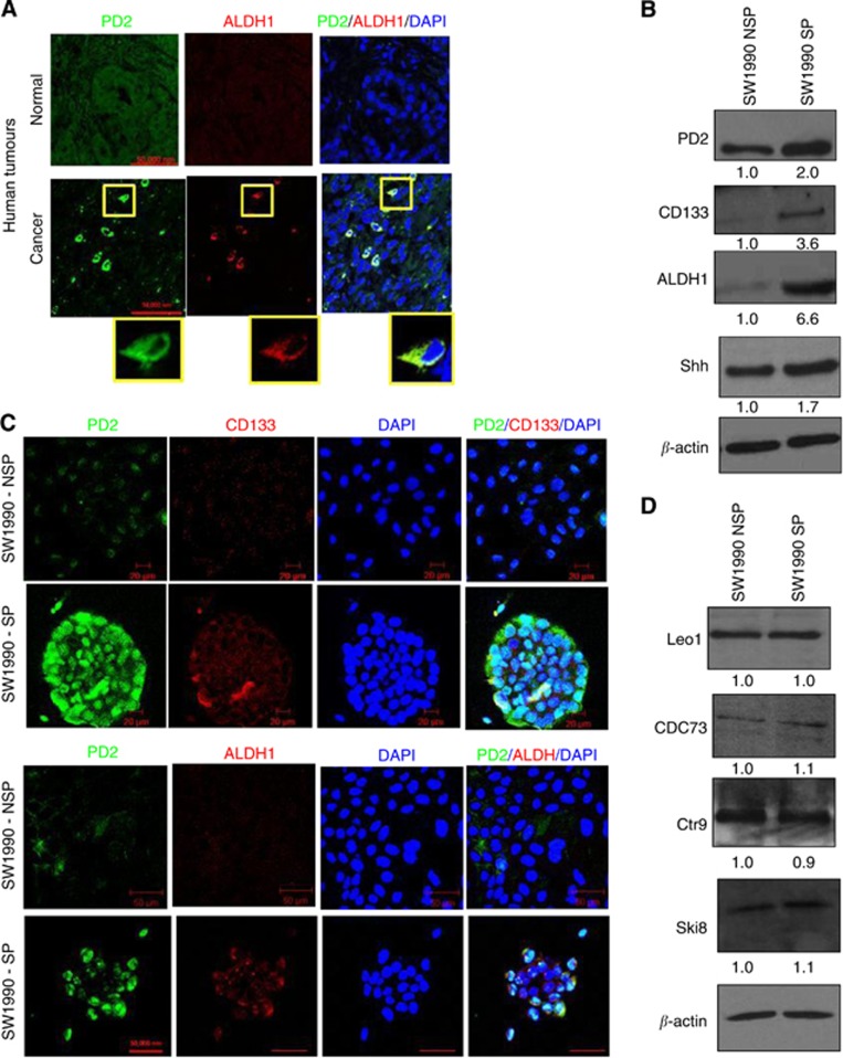
Figure 3.
Expression of PD2 and CSC markers. SP and NSP cells were processed for protein extraction and western blotting using standard procedures. (A) Overexpression of PD2 in human pancreatic tumours along with ALDH1 in specific cells. Control tumours did not show specific overexpression of PD2. (B) Western blot analysis showed increased expression of PD2 in isolated SP cells along with cancer stem cell-specific markers (CD133 and ALDH1) and also the self-renewal marker SHH in SW1990-SP cells compared with NSP cells. β-actin was used as a loading control. Fold change of band intensity is mentioned in the western blot results. (C) Confocal analysis showed increased expression of PD2 (green) along with CSC markers (CD133 and ALDH1, red) in SP cells compared with NSP cells (DAPI-nuclear staining). (D) Western blot analysis showed other PAF complex subunits such as Leo1, Cdc73, Ctr6 and Ski8 did not show any variation of expression in both SP and NSP cells.

