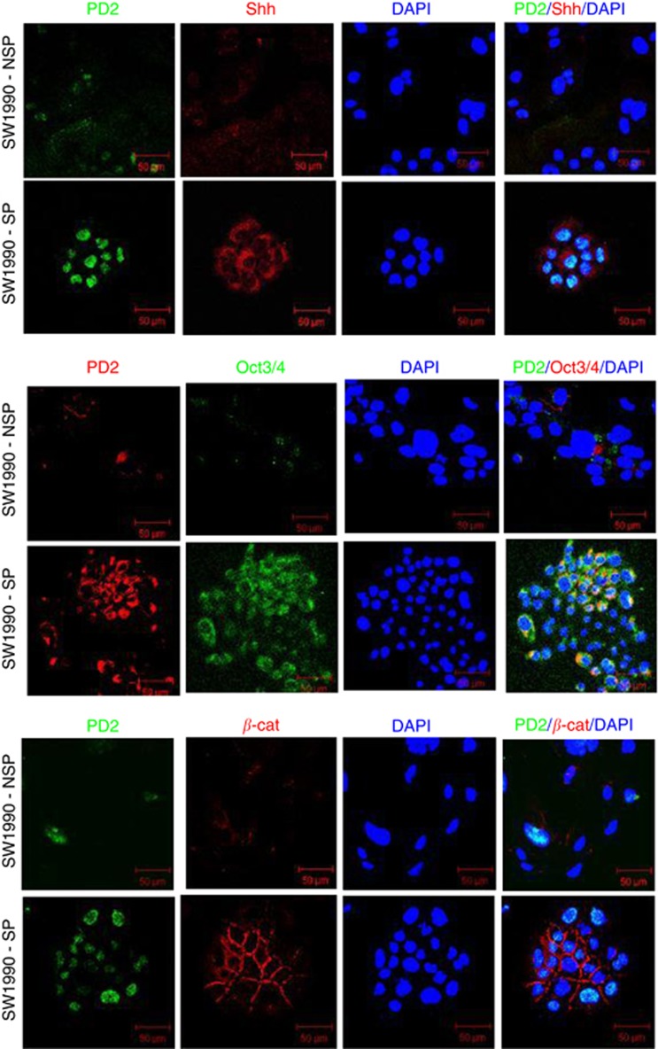
Figure 4.
Self-renewal markers in isolated SP cells. Confocal analysis showed a set (Shh, Oct3/4 and β-catenin) of self-renewal marker expression in SW1990-SP cells. The first panel showed the increased expression of Shh (red) along with PD2 (green) in SP cells compared with NSP cells. The second panel showed the increased expression of Oct3/4 (green) along with PD2 in SP cells compared with NSP cells. The third panel showed the membrane localisation and increased expression of β-catenin (red) along with PD2 (green) expression. DAPI was used for nuclear staining.

