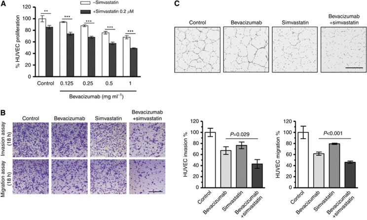
Figure 1.
Simvastatin directly inhibits HUVEC viability, migration, invasion, and tube formation in vitro. (A) Cell viability assay of HUVECs treated with 0.2 μM simvastatin in addition to various doses of bevacizumab (0, 0.125, 0.25, 0.5, or 1 mg ml−1). At 24 h after treatment, cells were stained with fluorescent dye (1 μM calcein-AM) for 30 min. Results of the cell count are expressed as the percentage of cell viability using the control PBS as a reference (**P=0.003, ***P<0.001). (B) Invasion and migration assays using a double chamber method. A total of 1 × 10⁵ colon cancer cells were seeded in a 24-well plate (the lower chamber) and cultured overnight, and HUVECs (5 × 10⁴ cells) treated with or without 0.2 μM simvastatin or 0.25 mg ml−1 bevacizumab were seeded in Matrigel-precoated transwell chambers. Transwell chambers were then placed in 24-well plates. After 24-h incubation, upper surfaces of transwell chambers were wiped with a cotton swab, and invading cells were fixed and stained with a 0.05% (w/v) crystal violet solution. Results of the cell count are expressed as the percentage of cell invasion using the control PBS as a reference. Addition of simvastatin to bevacizumab treatment induced a statistically significant decrease in HUVEC invasion (HUVEC invasion, P=0.029; HUVEC migration, P<0.001). (C) Tube formation assay of HUVECs treated with or without 0.2 μM simvastatin or 0.25 mg ml−1 bevacizumab. The ECM gel was thawed at 4 °C overnight and then bottom coated in a 96-well plate at 37 °C for 30 min. Next, 150 μl of media containing HUVECs (1 × 10⁶ cells) treated with or without 0.2 μM simvastatin or 0.25 mg ml−1 bevacizumab was added to each well on top of the solidified ECM gel and incubated at 37 °C for 18 h. Tubes were subsequently stained with the fluorescent dye (1 μM calcein-AM) for 30 min (size bar, 400 μm).

