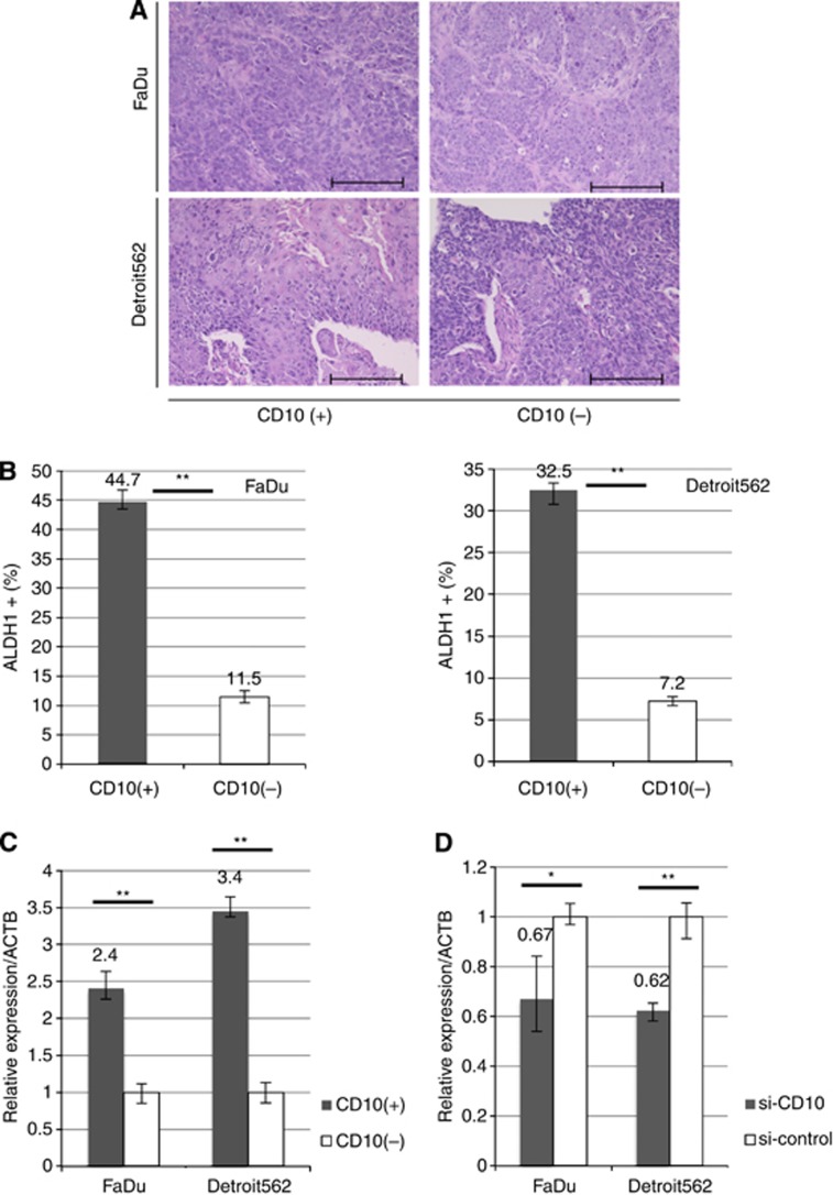
Figure 4.
Histology of tumours from CD10(+)/(–) subpopulations and the relationship between CD10 and other stem cell markers. (A) H&E staining of FaDu and Detroit562 xenograft tumours. Scale bar, 100 μM. (B) Expression of ALDH1 in CD10(+)/(−) FaDu and Detroit562 cells was assessed by FACS. Data represent means±s.e.m.; **P<0.01. (C) OCT3/4 expression in CD10(+)/(−) FaDu and Detroit562 cells was assessed by qRT–PCR. (D) OCT3/4 expression in FaDu and Detroit562 following transfection with either si-CD10 or si-control was assessed by qRT–PCR. Gene expression levels are presented as a ratio of the internal control, ACTB±s.e.m. *P<0.05; **P<0.01.

