Figure 2.
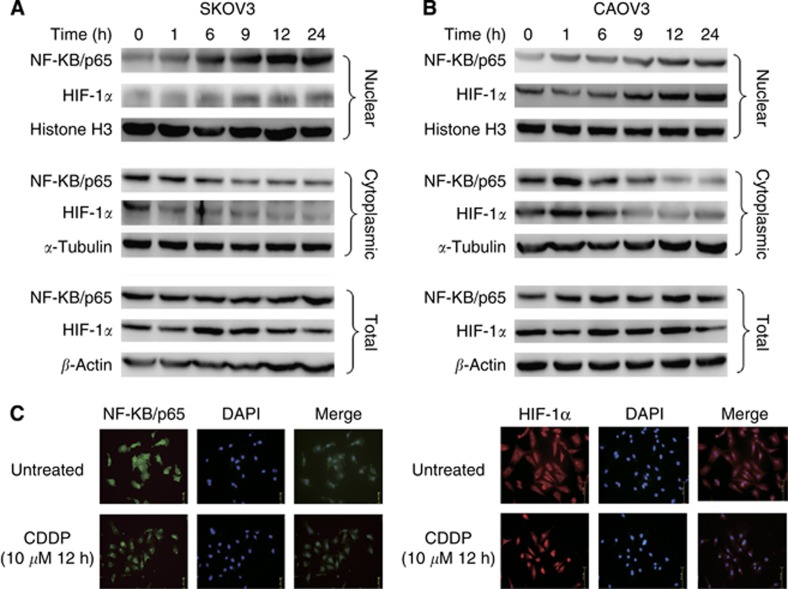
Treatment with CDDP enhances the nuclear localisation of NF-κB/p65 and HIF-1α. The SKOV-3 (A) and CAOV-3 (B) cells were treated with CDDP (10 μM) at different time intervals (0–24 h). Thereafter, nuclear, cytoplasmic and total extracts were prepared and the expression and localisation of NF-κB/p65 and HIF-1α were examined by immunoblot analysis. Histone H3 (for the nuclear fraction), α-tubulin (for the cytoplasmic fraction) and β-actin (for the total fraction) were used as loading controls. (C) The localisation of NF-κB/p65 and HIF-1α in SKOV-3 cells with or without 10 μM CDDP for 12 h treatment were analysed using immunofluorescence. The data indicate increased levels of NF-κB/p65 and HIF-1α in the nuclei of the ovarian cancer cells treated with CDDP.

