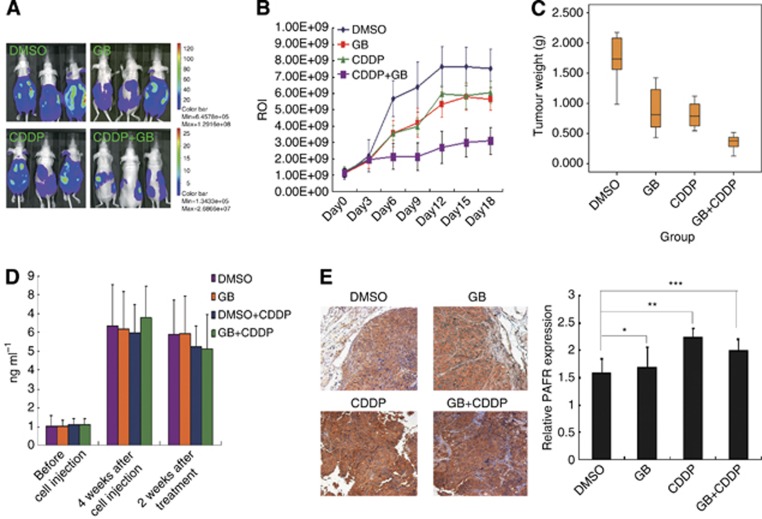
Figure 7.
Inhibition of PAFR significantly sensitises CDDP therapy in an ovarian cancer model. (A) The representative in vivo imaging picture of the four groups of nude mice with peritoneal ovarian cancer. SKOV-3-luc cells were intraperitoneally injected into mice and the ROI was determined. (B) The ROI values represented the tumour size in each group as measured by a luciferin imaging system every 3 days. (C) Boxplots showing the tumour weight in four groups at the end of therapy. (D) The concentrations of mouse serum PAF were determined in each of the four groups before and after treatment with the different drugstore combinations. (E) Protein expression of PAFR in ovarian cancer tissues of four groups by immunohistochemistry (IHC). After 3 weeks of CDDP treatment, the PAFR expression was increased significantly (IHC, 10 × , *P>0.05, **P<0.05, ***P<0.01). CDDP, CDDP treatment group; DMSO, DMSO treatment group; GB, GB treatment group; GB+CDDP, GB+CDDP treatment group.

