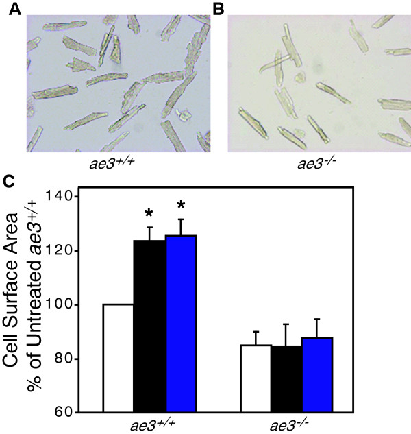
Figure 4.

Effect of hypertrophic stimuli on cardiomyocyte size. Cardiomyocytes isolated from the ventricles of WT (ae3+/+)(A) and ae3−/−(B) adult mice were subjected to vehicle alone (control), ANGII (1 μM) and PE (10 μM) treatment for 24 h, following an 18 h pre-treatment period. Images of the cardiomyocytes, taken pre- and post-treatment using a QICAM fast cooled 12-bit color camera, were quantified to measure the cell surface area. In the control group, equal volume of the vehicle was added. C, Cell surface areas were expressed as a percentage of vehicle-treated control groups (open bars) and compared to the ANGII (black bars) and PE (blue bars) treated groups. *P < 0.05, relative to control group (n = 10 mice in each group).
