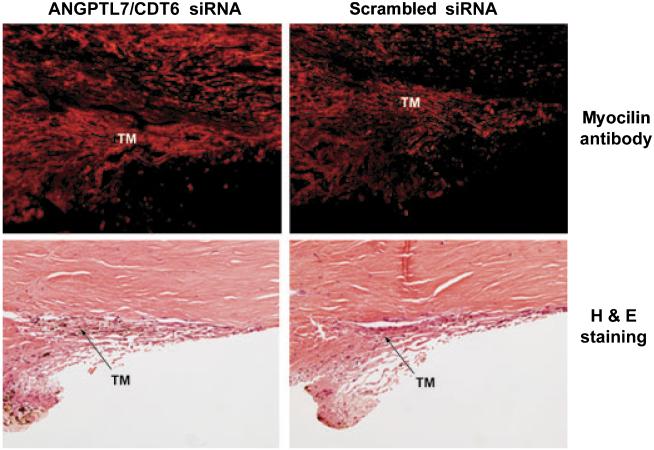
Figure 7.
Histologic assessment of human trabecular meshworks from anterior segments perfused with ANGPTL7/CDT6 and Scrambled siRNA. Top: MYOC expression examined by immunohistochemistry on OCT-embedded frozen sections from blocks containing tissue wedges from opposite quadrants of eye pair #1. Ten-micrometer sections were stained with goat anti-human MYOC and donkey anti-goat Alexa Fluor 594. Bottom: 5-lm paraffin sections from opposite quadrants of the same eye pair stained with H&E. Quantification of fluorescence intensity showed that MYOC expression is higher in the eye perfused with ANGPTL7/CDT6 siRNA than in the contralateral eye perfused with Scrambled siRNA. Although the architecture of the trabecular meshwork is well conserved in both eyes, the eye perfused with ANGPTL7/CDT6 siRNA appears to have more extracellular matrix that tends to constrict the Schlemm’s canal.

