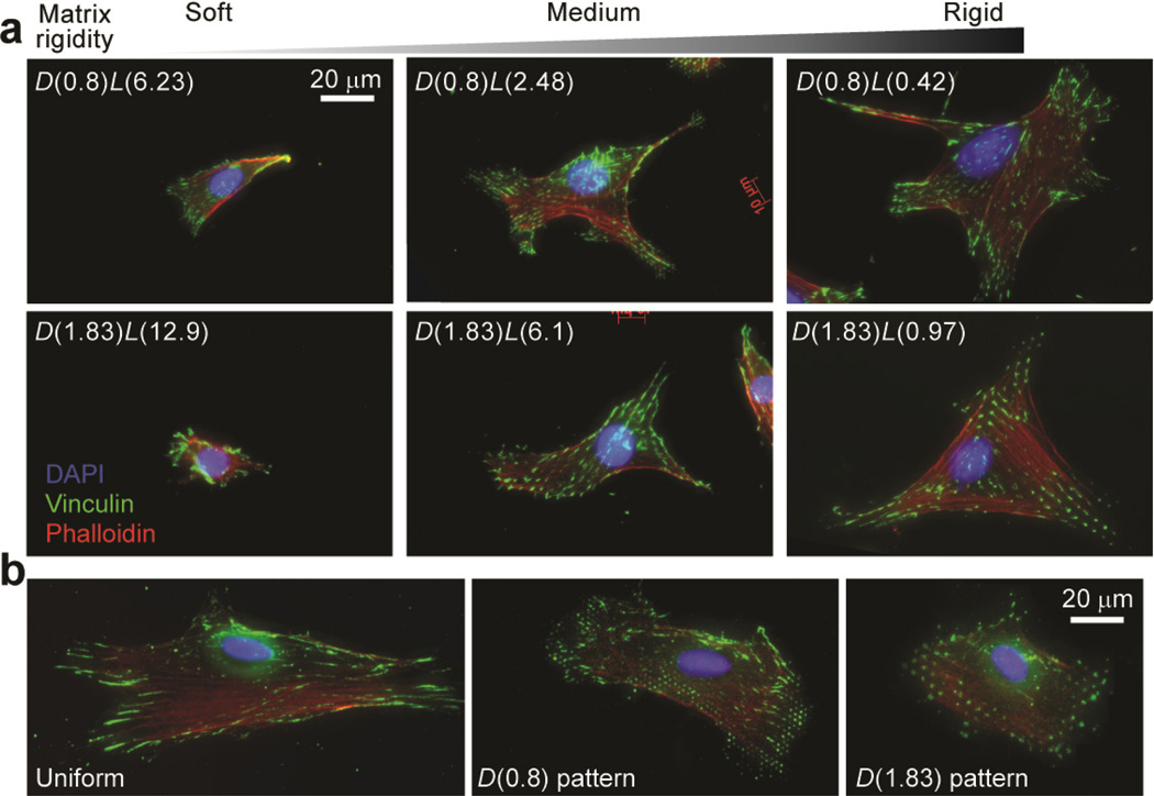
Figure 3.
Representative immunofluorescence images of single NIH/3T3 cells plated on different PDMS-based substrates. Cells were grown overnight in the complete growth medium prior to fixation, and were stained with DAPI, fluorophore-labeled phalloidin and anti-vinculin to visualize nuclei, actin filaments, and FAs, respectively. (a) Single NIH/3T3 cells plated on the PDMS micropost arrays of different geometries and rigidities as indicated. (b) Single NIH/3T3 cells plated on flat PDMS surfaces coated with different ECM patterns as indicated.

