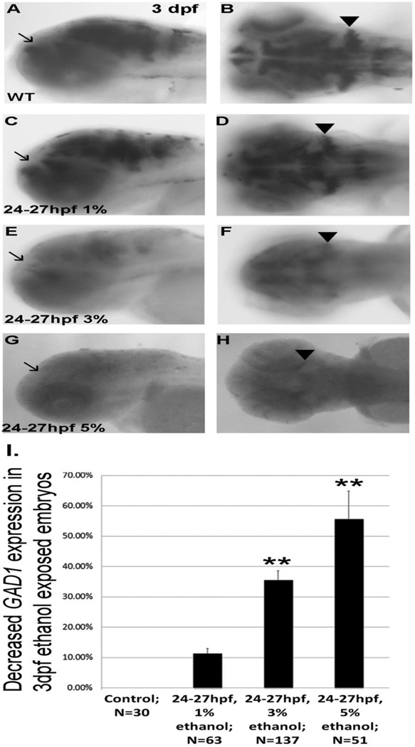
Figure 7.
GAD1 expression in 3 dpf forebrain and hindbrain following transient binge-like ethanol exposure. A, B, lateral and dorsal views of embryos not exposed to ethanol; C, D, lateral and dorsal view of embryos exposed to 1% ethanol from 24–27 hpf; E, F, lateral and dorsal view of embryos after 3% ethanol; G,H, lateral and dorsal view of embryos following 5% ethanol. Arrows denote forebrain expression of GAD1, and arrowheads indicate expression of GAD1 in cerebellum. I, quantitation of decreased GAD1 expression in ethanol exposed embryos. Data are shown as Mean ± SD for at least 3 independent experiments, with total number of embryos analyzed shown. **, significantly different from control GAD1 expression, P< 0.0001.

