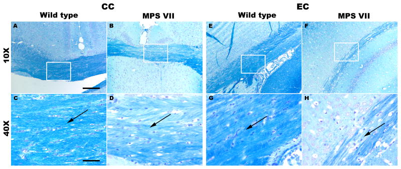
Figure 5.
(A-H) Luxol fast blue (LFB)-stained brain sections from the CC (A-D) and EC (E-H) regions of the brain showing myelin staining in wild type (A, C, E, G) and mucopolysaccharidosis type VII (MPS VII) (B, D, F, H) mice. The rectangular boxes on sections at magnification of 10x (scale bar = 200 μm) are zoomed at 40x (scale bar = 50 μm) (C, D, G, H). Black arrows indicate splitting of the fibers and degree of compactness of the white matter fiber bundles in wild-type and MPS VII mice. CC, corpus callosum; EC, external capsule.

