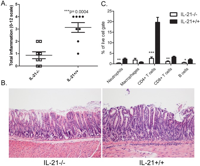
FIG 2 .
Inflammation is reduced in H. pylori-infected IL-21−/− mice compared to the levels in their H. pylori-infected wild-type littermates. Levels of acute and chronic inflammation were scored for stomach tissue (in the corpus and antrum) at 3 months postinfection. (A) Total inflammation was scored on a scale of 0 to 12 (bars represent means, and error bars represent upper and lower interquartile ranges). (B) Representative sections of the gastric mucosa from 3 months postinfection are presented (×200). (C) Flow cytometric analysis was performed on dissociated stomach tissue at 3 months postinfection (n = 8 per genotype). Percentages of neutrophils (Gr1+ CD11c+), macrophages (CD11b+ Gr1−), CD4+ CD3+ T cells, CD8+ CD3+ T cells, and B cells (B220+) were calculated in the live-cell gate from the H. pylori-infected mice. Bars and error bars represent means ± SEM. *, P ≤ 0.05; **, P ≤ 0.01; ***, P ≤ 0.001. Results are representative of 3 independent experiments.

