Figure 3.

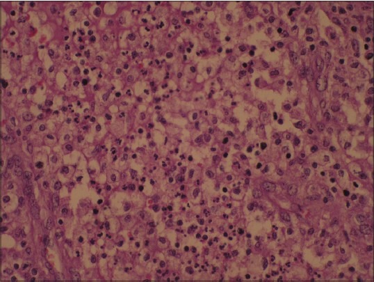
Microphotograph showing sheets of foamy cells with normochromic nuclei, admixed with mixed inflammatory cells (H and E, ×400)

Microphotograph showing sheets of foamy cells with normochromic nuclei, admixed with mixed inflammatory cells (H and E, ×400)