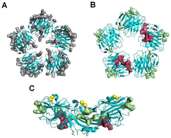
Figure 9. Identification of a novel Fcγ receptor binding site on SAP.
A) Mutated amino acid residues are indicated by molecular surface representation (gray) on the SAP structure. B) The functionally significant amino acid residues (green) are distinct from the previously identified FcγRIIa binding site (red). C) When E153 is excluded; the remaining functionally significant amino acid residues form a distinct binding site. The yellow spheres represent the two calciums bound to SAP.

