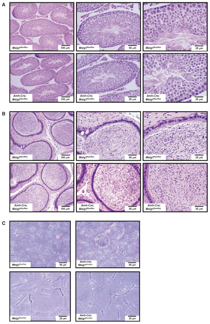
Figure 7.

Disruption of Meig1 gene in the Sertoli cells does not affect normal spermatogenesis. (A) Representative testicular histology from a Meig1flox/flox mouse and a Amh-Cre; Meig1flox/flox mouse. Both reveal normal architecture of the seminiferous tubules and interstitial tissue. (B) Representative epididymides histology from a Meig1flox/flox mouse and a Amh-Cre; Meig1flox/flox mouse. The lumen is filled with mature spermatozoa in both mice. (C) Morphology of epididymal spermatozoa collected from a Meig1flox/flox mouse and a Amh-Cre; Meig1flox/flox mouse. All spermatozoa in both groups appear to be normal.
