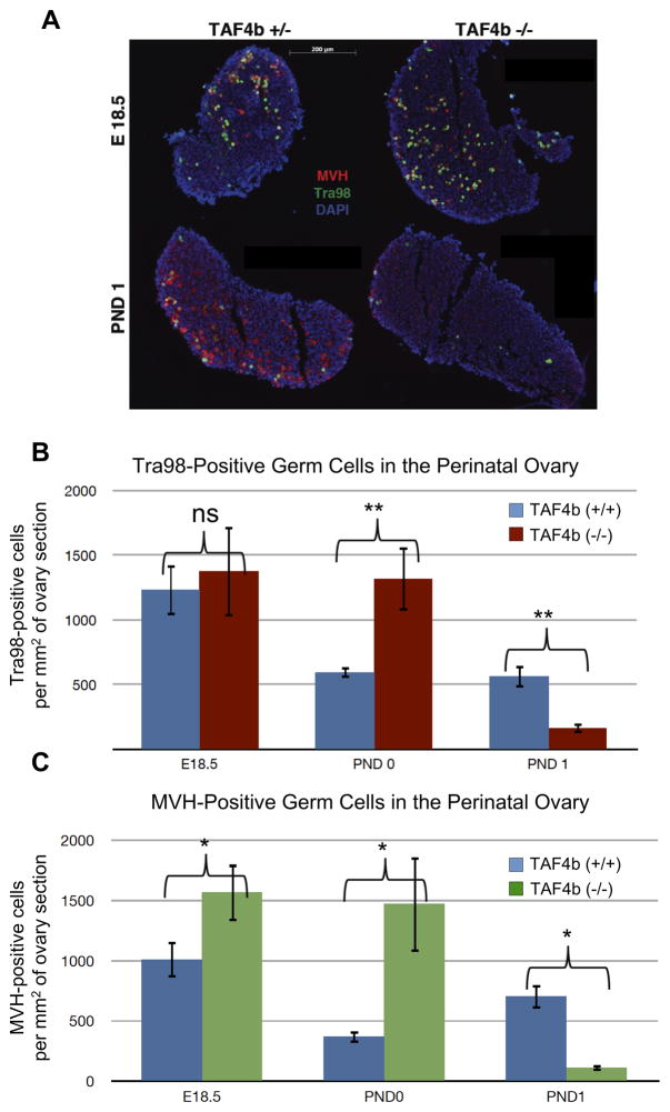
Figure 1. Perinatal oocyte depletion in TAF4b (−/−) ovaries.
A) MVH (red) and Tra98 (green) staining were used as cytoplasmic and nuclear germ cell markers, respectively, in 8μm ovary tissue sections. DAPI (blue) denotes nuclei. TAF4b (+/−) and (−/−) ovaries exhibit comparable germ cell densities at E 18.5 (top), while TAF4b (−/−) ovaries suffer excessive depletion of oocyte by PND 1 (bottom). B) Quantification of stained tissue sections. Tra98-positive oocytes were counted per section and total number was divided by total DAPI area. N=4 for PND 0 TAF4b (+/−) and (−/−). N=5 for E18.5 TAF4b (−/−). N=6 for E18.5 TAF4b (+/−), PND 1 TAF4b (+/−), and PND 1 TAF4b (−/−). **: two-tailed T-test, p<0.01. C) Independent quantification of stained tissue sections. MVH-positive oocytes were counted per section and total number was divided by total DAPI area. N=3 for all groups. *: two-tailed T-test, p<0.05.

