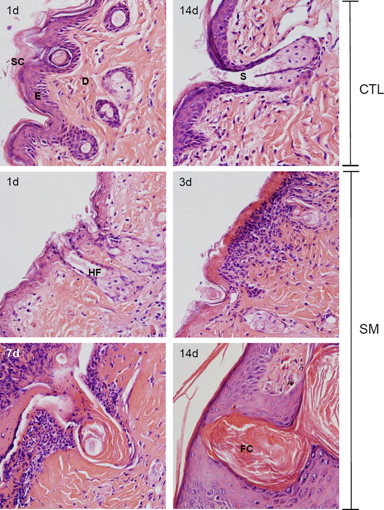
Figure 1. Structural changes in hairless mouse skin following SM.
Histological sections prepared 1 and 14 days after air exposure (CTL), and 1, 3, 7 and 14 days after SM exposure were stained with H&E. One representative section from 3 mice/treatment group is shown (original magnification, × 400). Stratum corneum (SC), epidermis (E), hair follicle (HF), dermis (D), sebaceous gland (S), follicular cyst (FC).

