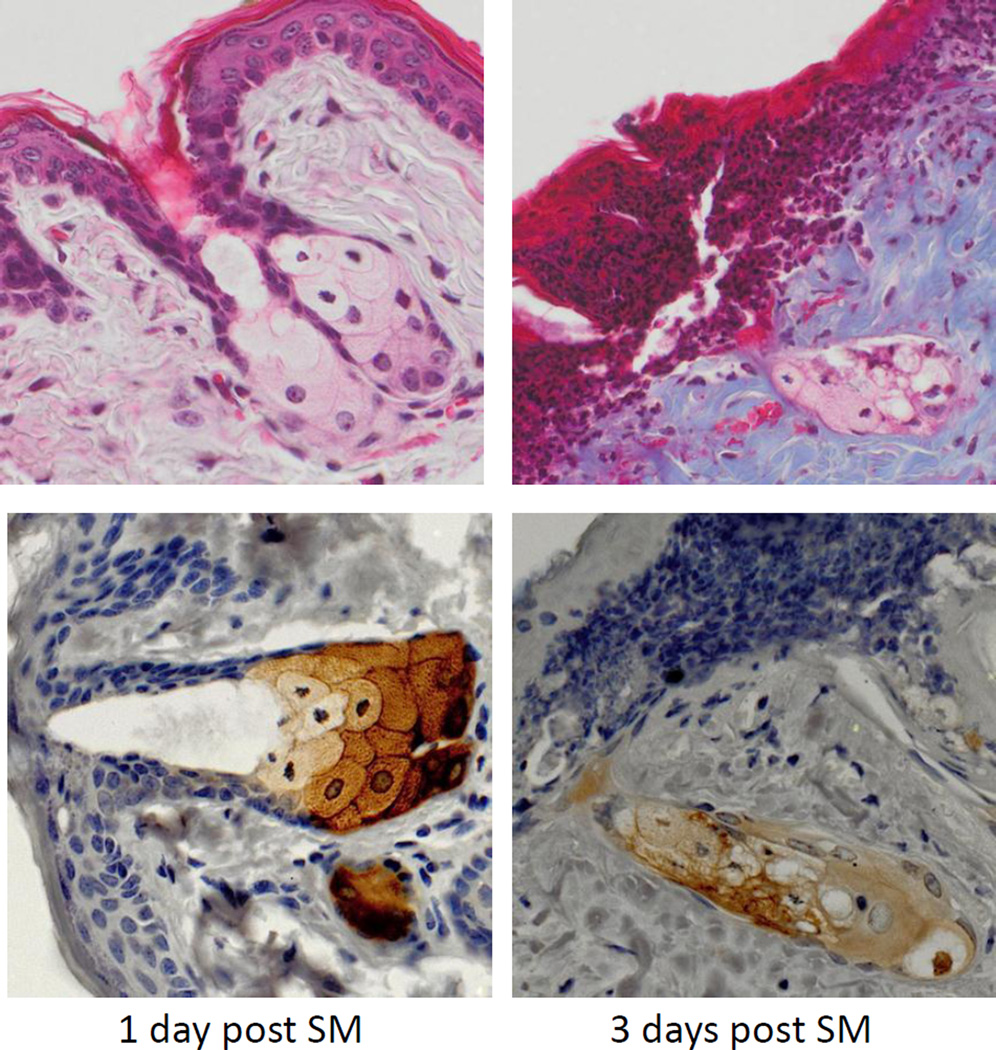
Figure 4. Effects of SM on sebaceous glands.
Histological sections prepared 1 day and 3 days after SM exposure were stained with Gomori’s trichrome followed by aniline blue counterstaining (upper panels) or with anti-fatty acid synthase antibody (lower panels). One representative section from 3 mice/treatment group is shown (original magnification, × 400).

