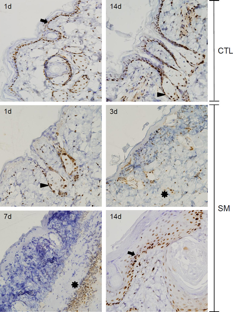
Figure 7. Effects of SM on PCNA expression.
Histological sections prepared 1 and 14 days after air exposure (CTL), and 1, 3, 7 and 14 days after SM exposure were stained with an antibody to PCNA. Antibody binding was visualized using a Vectastain Elite ABC kit (original magnification, × 400). One representative section from 3 mice/treatment group is shown. Arrows indicate interfollicular and outer root sheath basal keratinocytes, arrowheads indicate sebocytes and asterisks indicate inflammatory cells at the dermal/epidermal junction.

