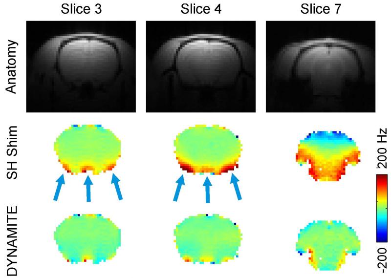
Fig. 4. DYNAMITE VS. SH SHIMMING FOR EPI.
Comparison of SH and DYNAMITE magnetic field shimming. First row: Standard gradient-echo structural images of the rat brain. Center row: Localized field imperfections of varying polarity remain in the rat brain at 11.7 Tesla after static (global) first through third order spherical harmonic (SH) shimming, since the complexity of the distortions exceeds the modeling capability of SH field shaping. Third row: The combination of MC field modeling and a dynamic approach for shimming enables largely improved magnetic field homogeneity throughout the brain.

