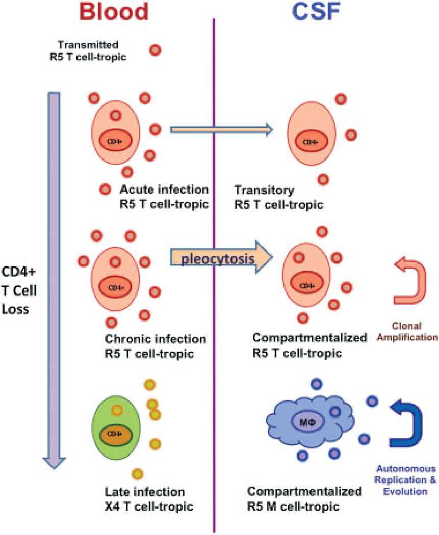
Fig. 3.
Model of types of central nervous system (CNS) infection reflected in cerebrospinal fluid (CSF). Systemic infection is shown on left and CNS on right. Acute and chronic infection by T-tropic, CCR5-using (R5) viruses generally derive from circulating systemic virus populations and likely relate to transport into the CSF by CD4+ T cells with limited or short-lived local amplification. Only occasionally do these undergo clonal amplification, one form of compartmentalization, and rarely do they further evolve and diversify locally. This contrasts to macrophage- (M-) tropic HIV-1 populations, which do not require high CD4 levels to infect cells and can propagate within the CNS in macrophages and related cells—comprising the second and more pathogenic type of compartmentalized infection associated with HIV encephalitis and HIV-associated dementia HAD. The location and events underlying emergence of M-tropic viruses in the CNS remain uncertain. An independent evolution occurs systemically with the emergence of T-tropic CXCR4-using (X4) viruses systemically in some patients; these are associated with low-blood CD4 cells and rapid progression, and may be found in CSF. However, there is no evidence that they are directly neuropathic. In fact, R5 viruses continue to predominate in patients with low CD4 cells, but are not included in the bottom portion in this crowded figure. The figure derives from concepts developed by Swanstrom and colleagues in their work on CSF-virus phylogeny and tropism.30

