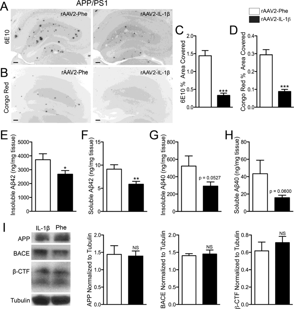
Figure 3.
rAAV2-IL-1β reduces amyloid deposition in 7 mo old APP/PS1 mice without altering APP or processing. A,B, Representative images of 6E10 (A) and Congo red (B) staining of amyloid plaques in APP/PS1 mice transduced with either rAAV2-Phe or rAAV2-IL-1β. Scale bar = 30 µm. C,D, Quantification of 6E10 (C) and Congo red (D) displayed as percent area of hippocampus covered by amyloid plaques. E,F,G,H, ELISA measurements of hippocampal levels of insoluble Aβ1-42 (E), soluble Aβ1-42 (F), insoluble Aβ1-40 (G), and soluble Aβ1-40 (H). n = 9-12 per group. Data displayed as mean ± SEM, unpaired t-test, *p < 0.05, **p < 0.001, ***p < 0.0001. I, Representative Western blot images and quantification of band intensities for APP, BACE, and β-CTF normalized to tubulin. n = 4-9 per group. Data displayed as mean ± SEM, Mann-Whitney test.

