
Fig. 1.
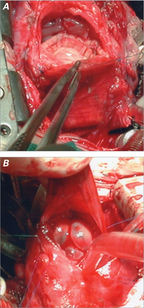
A) Intraoperative photograph shows the BioPulmonic valve injected into the right ventricular outflow tract. The enlarged tract is seen from the cephalic perspective. The enlargement patch has been sutured onto the pulmonary trunk, and the infundibular suture is yet to be completed. B) The same valve is shown from the caudal perspective. The prosthetic cusps are closed.