
Fig. 4.
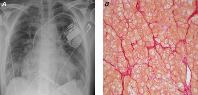
November 2012. At the time of the 2nd left ventricular assist device implantation, A) chest radiograph shows cardiomegaly with pulmonary congestion, and B) photomicrograph shows mild-to-moderate hypertrophy with moderate perivascular and interstitial fibrosis (picrosirius red stain, orig. ×10). The hypertrophy is comparatively reduced, and the histologic appearance is similar to the findings before the first implantation (see Fig. 2A). The interstitial fibrosis appears to have increased only minimally since the first device was explanted (see Fig. 3B).