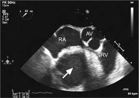
Fig. 3.

Patient 2. Transesophageal echocardiogram (short-axis view) at the level of the aortic valve shows a large, homogeneous, echogenic mass (arrow) compressing the right atrium and right ventricle.
AV = aortic valve; RA = right atrium; RV = right ventricle
